

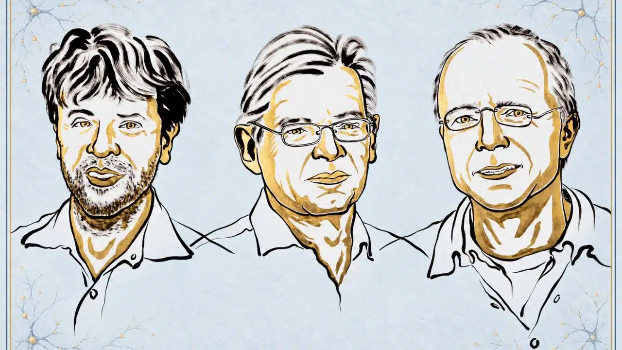
The 2026 Nobel Prize in Physiology or Medicine was awarded to Karl Deisseroth, Peter Hegemann and Georg Nagel for their work on optogenetics, a method that lets scientists control cells with light. These three scientists turned a light-sensitive protein into a way to test what nerve cells do. Their work has transformed brain research, while its medical promise is only beginning to be tested in patients.
The Nobel committee’s first call to Karl Deisseroth came at 12:27 a.m. in California. He missed it. Seconds later, his wife’s phone rang. The Nobel Prize in Physiology or Medicine 2026 had been awarded to Deisseroth, Peter Hegemann and Georg Nagel for discoveries that let scientists control selected nerve cells with light.
The announcement, made on October 5, recognizes the development of optogenetics. It is a powerful research method with an unlikely starting point: a tiny alga that swims toward light. By working out how the alga senses light, and then adapting that mechanism for nerve cells, the laureates helped researchers investigate how activity in the brain shapes movement, sleep and behavior.
The Nobel Assembly honored them “for their discoveries concerning light-gated ion channels and optogenetics.” Deisseroth is a professor at Stanford University and an investigator with the Howard Hughes Medical Institute. Hegemann works at Humboldt University of Berlin, and Nagel at the University of Würzburg. They will share a prize of 12 million Swedish kronor.
The finding deserves its place in medicine even though optogenetics is chiefly a laboratory tool today. It has changed what scientists can test about the brain. Efforts to turn that knowledge into treatments are further along in the eye than in the brain, and the difference matters.
The clue came from an alga
Hegemann wanted to understand how Chlamydomonas, a single-celled alga, detects light quickly enough to swim toward it. Working with Nagel and other colleagues, he helped uncover proteins called channelrhodopsins.
A channelrhodopsin sits in a cell’s outer membrane. When light of the right wavelength reaches it, the protein opens a passage for electrically charged particles, or ions. Their movement changes the cell’s electrical state. In 2003, Nagel, Hegemann and colleagues showed that one of these proteins, channelrhodopsin-2, could make other kinds of cells respond directly to light.
That result offered an answer to a long-standing problem in neuroscience. Nerve cells communicate through electrical signals, but most do not respond to light on their own. If researchers could give a chosen group of neurons the gene for a light-sensitive protein, they might be able to activate those cells with a precisely timed flash.
The idea sounds simple now. Making it work meant getting the protein into the right neurons, ensuring it reached the cell surface, and delivering light to those cells without overwhelming the surrounding tissue.
From a light-sensitive cell to a living brain
Making a nerve cell respond to light was only the first step. Scientists then had to target specific neurons and reach them inside a living brain. Once they could do that, they could test whether activity in those cells changed an animal’s behavior.
The 2005 neuron experiment
In a landmark 2005 experiment, Deisseroth and colleagues showed that blue light could trigger electrical activity in cultured rat nerve cells carrying channelrhodopsin-2. The published paper had five authors: Edward Boyden, Feng Zhang, Ernst Bamberg, Nagel, and Deisseroth.
Researchers subsequently combined genetic targeting with ways to deliver light into the brains of living animals. Other light-sensitive proteins expanded the toolkit, allowing scientists to suppress some neural activity as well as stimulate it. These are different molecular tools; channelrhodopsin-2 itself is primarily used to activate cells.
A way to test cause and effect
That combination of genes and light is the basis of optogenetics. Its value lies in timing and selectivity. Brain imaging can show that an area is active during an experience. Optogenetics lets a researcher change the activity of a defined group of cells and observe what follows.
A 2007 study offers a clear example. Scientists used light to stimulate a particular class of neurons involved in arousal in sleeping mice. The stimulation increased the chance that the animals would wake up. The experiment connected activity in those cells to a change in behavior. It did not reduce the whole experience of sleep to a single switch.
A change in behavior after stimulation is evidence about a circuit under particular experimental conditions. It is not, by itself, a complete explanation of a human feeling or a disease. Still, the ability to test a proposed connection so directly has reshaped brain research.
The first steps toward treating people are in the eye
The eye presents an opportunity that the brain does not. Light already enters it, and some retinal cells can survive after the cells that normally detect light have died. In inherited disorders such as retinitis pigmentosa, researchers are testing whether they can make surviving cells light-sensitive and recover some useful visual function.
What one patient could see in 2021
In 2021, a research team reported partial visual recovery in one person with advanced retinitis pigmentosa. They delivered the gene for a light-sensitive protein to cells in his retina and used special goggles to project light patterns into the treated eye.
With the goggles, he could perceive and locate some objects during tests. Without them, the reported improvement was not seen. It was a demonstration of possibility, not a return to ordinary vision.
A different approach tested in four people
The work has moved beyond that single case, though different teams are testing different approaches. A study published in 2025 described four people with severe retinitis pigmentosa who received an experimental treatment called MCO-010. It uses an engineered light-sensitive protein intended to work with ambient light and targets a different layer of retinal cells from the 2021 treatment.
The researchers reported improvements on measures of vision and mobility during follow-up. Four people are too few to establish how well a treatment works across patients. The study also had no comparison group, and individual results varied. Its findings support further testing; they do not prove that the treatment reliably restores sight.
A larger trial reaches regulatory review
A separate trial of MCO-010 enrolled 27 participants, including nine assigned to a sham procedure. Its developer, Nanoscope Therapeutics, says the trial met its principal vision measures. That is the company’s account of the results, which remain subject to regulatory assessment.
In September 2026, Nanoscope announced that the US Food and Drug Administration had accepted its application to review MCO-010 for people with severe vision loss from retinitis pigmentosa. The company also announced that a related application entered priority review in Japan on October 1.
Acceptance for review is not approval. Regulators still have to assess the evidence on benefit, safety and how long any improvement lasts. A useful result for a patient may mean finding an object or navigating a space more easily; it should not be described as the restoration of normal sight.
Nobel Prize in Physiology or Medicine: What the Nobel recognizes
The three laureates represent different stages of the discovery. Hegemann’s investigation of how algae sense light and Nagel’s work establishing the properties of channelrhodopsins supplied the molecular foundation. Deisseroth and his collaborators made light control of neurons a practical way to study brain circuits.
The papers also show how many people contributed. The Nobel Prize can be divided among no more than three individuals, while the experiments that established optogenetics involved wider teams. Reading the award alongside those papers gives a fuller picture of the work without diminishing the discoveries being honored.
For me, this is the most convincing reason the Nobel matters: optogenetics changed the questions scientists could answer. A method that reveals whether a particular cell population contributes to a response has lasting value even before it produces a treatment. That is also why the clinical claims now associated with the field need to be judged on their own evidence.
Why the brain is a harder destination
Progress in the retina cannot be assumed to carry over to depression, Parkinson’s disease or other brain disorders. Light can reach the eye naturally. Reaching a specific circuit deep inside the brain generally requires a more invasive way to deliver it. Researchers would also need to introduce a light-sensitive protein safely into the intended cells and understand the lasting effects of doing so.
The biology presents a second challenge. A circuit that changes an animal’s behavior in an experiment may be one part of a much larger process. Human brain disorders vary between people and involve networks of cells, not a single faulty switch. Optogenetic studies can help reveal those networks, but they do not yet amount to established optogenetic treatments for brain disease.
That gap should shape how we read the Nobel Prize news. The discoveries are already consequential because they gave researchers an unusually precise way to investigate living circuits. Retinal trials are now testing whether a related idea can provide meaningful benefits to patients. Treatments aimed at the human brain remain a longer and more difficult prospect.
The story began with scientists asking why an alga swims toward light. More than two decades later, that question has helped them ask far sharper questions about the brain. The next measure of progress will be equally concrete: what people can safely do after treatment that they could not do before.