
Thyroid cancer is one of the most common endocrine malignancies, with an increasing incidence worldwide. While the majority of thyroid nodules are benign, distinguishing between benign and malignant nodules is crucial for appropriate management. Traditional ultrasound is a valuable tool in this assessment, but it has limitations in differentiating benign from malignant nodules. This is where ultrasound elastography for Thyroid cancer comes into play—a relatively new imaging technique. It offers a non-invasive method to evaluate the stiffness of thyroid nodules, potentially improving the diagnostic accuracy of thyroid cancer.
Understanding Thyroid Cancer
Thyroid cancer originates in the thyroid gland, a butterfly-shaped organ located at the base of the neck. The thyroid gland produces hormones that regulate metabolism, heart rate, and body temperature. Thyroid cancer can present as a nodule or lump in the thyroid, and while many nodules are benign, a small percentage are cancerous. The main types of thyroid cancer include:
- Papillary thyroid cancer: The most common type, known for its slow growth and high survival rate.
- Follicular thyroid cancer: Less common and more likely to spread to other parts of the body.
- Medullary thyroid cancer: Arises from C cells in the thyroid and can be associated with genetic syndromes.
- Anaplastic thyroid cancer: A rare, aggressive form of thyroid cancer that is difficult to treat.
The Role of Ultrasound in Thyroid Cancer Diagnosis
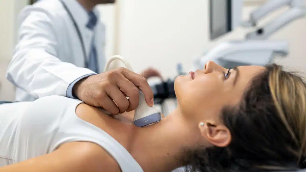
Conventional ultrasound is the first-line imaging modality used to evaluate thyroid nodules. It provides information on the size, shape, composition, and vascularity of the nodule. Certain ultrasound features, such as hypoechogenicity (darker appearance), irregular margins, microcalcifications, and increased blood flow, are associated with a higher risk of malignancy. However, these features are not definitive, and there is significant overlap between benign and malignant nodules. As a result, many patients undergo fine-needle aspiration biopsy (FNAB) to obtain a tissue diagnosis. FNAB, while effective, is invasive and sometimes yields indeterminate results, leading to repeated biopsies or unnecessary surgery.
What is Ultrasound Elastography?
Ultrasound elastography is an advanced imaging technique that measures tissue stiffness by assessing the mechanical properties of the tissue. The principle behind elastography is that malignant tissues tend to be stiffer than benign tissues. Elastography can be performed using different methods, with two main types being used for thyroid evaluation:
- Strain Elastography: Measures tissue deformation (strain) in response to an external force, such as the pressure applied by the ultrasound probe. Stiffer tissues deform less and appear darker on the elastogram, while softer tissues deform more and appear lighter.
- Shear Wave Elastography (SWE): Uses focused ultrasound waves to generate shear waves in the tissue. The speed of these waves is measured, with faster wave propagation indicating stiffer tissue. SWE provides a quantitative measure of tissue stiffness in kilopascals (kPa).
Benefits of Ultrasound Elastography in Thyroid Cancer Diagnosis
Ultrasound elastography offers several advantages in the evaluation of thyroid nodules:
- Non-invasive and Painless: Elastography is a non-invasive, painless procedure that can be performed alongside a standard ultrasound examination.
- Enhanced Differentiation: By providing information on tissue stiffness, elastography can help differentiate between benign and malignant nodules, potentially reducing the need for unnecessary biopsies.
- Quantitative Data: Shear wave elastography offers quantitative data on tissue stiffness, which can be useful in monitoring changes over time or in response to treatment.
- Real-Time Imaging: Elastography provides real-time imaging, allowing for immediate assessment of the nodule’s characteristics.
Clinical Applications of Ultrasound Elastography
- Risk Stratification of Thyroid Nodules:
Elastography can be used to assess the likelihood that a thyroid nodule is malignant. Nodules that are stiffer on elastography are more likely to be malignant. This information can be used alongside traditional ultrasound findings to stratify the risk of malignancy and guide the decision to perform a biopsy. - Guidance for Fine-Needle Aspiration Biopsy (FNAB):
Elastography can help target the stiffer areas of a nodule during FNAB, potentially increasing the diagnostic yield of the biopsy. This is particularly useful in nodules with heterogeneous composition, where the malignant area might be localized. - Follow-up of Thyroid Nodules:
For nodules that are not biopsied or are found to be benign, elastography can be used in follow-up assessments to monitor changes in stiffness over time. An increase in stiffness may warrant re-evaluation and possibly a repeat biopsy. - Assessment of Lymph Nodes:
Elastography can also be used to assess cervical lymph nodes in patients with known or suspected thyroid cancer. Stiffer lymph nodes on elastography are more likely to be metastatic.
Conclusion
Ultrasound elastography represents a significant advancement in the imaging and evaluation of thyroid nodules. By providing additional information on tissue stiffness, it enhances the ability to differentiate between benign and malignant nodules, potentially reducing the need for invasive procedures. While there are still challenges to be addressed, including operator dependency and the need for standardization, the future of elastography in thyroid cancer diagnosis looks promising.